Moscow Study proves AI cannot replace human emergency radiologists
The analysis showed that the best results were achieved when AI acted as a “second reader,” supporting physicians rather than replacing them.
Published
RUSSIA—Scientists in Moscow have found that the most effective way to diagnose hemorrhagic stroke from computed tomography (CT) scans is not through fully automated artificial intelligence (AI) systems, but through close collaboration between radiologists and AI tools.
The findings emerged from a large-scale study conducted by the Center for Diagnostics and Telemedicine of the Moscow Healthcare Department, which evaluated different approaches to integrating computer vision technologies into routine clinical practice.
Over a two-year period, specialists monitored the performance of three AI services and reviewed radiologists’ reports from more than 3,400 CT examinations conducted across 67 hospitals in Moscow.
The analysis showed that the best results were achieved when AI acted as a “second reader,” supporting physicians rather than replacing them.
In this model, the neural network rapidly identifies suspected bleeding sites, performs measurements, and presents preliminary findings, while the radiologist retains responsibility for the final diagnosis.
According to Yuri Vasilev, Medical Director of the Center for Diagnostics and Telemedicine, this collaborative approach significantly improves the detection of intracranial hemorrhages, including very small bleeds that can easily be overlooked.
Early identification is particularly important because timely treatment can reduce the risk of severe complications and improve patient outcomes.
AI supports, physicians decide
The study also revealed that radiologists continue to outperform AI systems when it comes to avoiding false-positive findings.
While AI can occasionally flag abnormalities that are not actually present, physicians are better able to distinguish genuine hemorrhages from imaging artifacts or other structures.
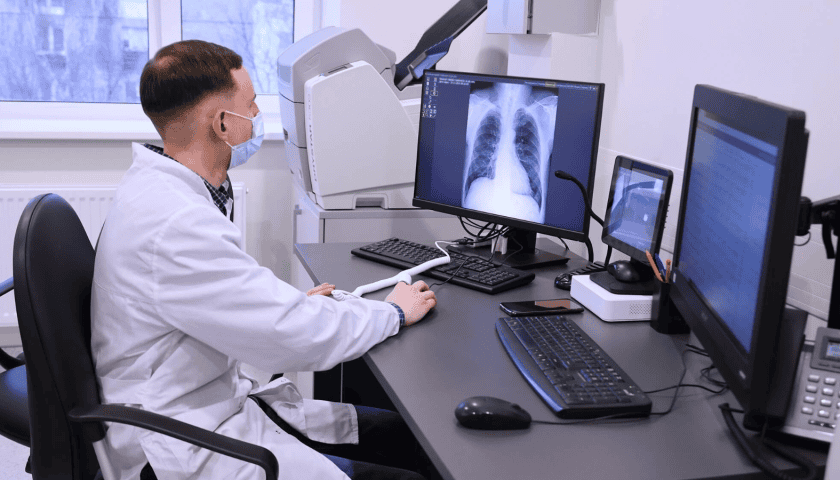
In practice, the workflow begins immediately after a CT scan is completed. The AI system receives the images, analyzes them within minutes, highlights suspicious regions, determines the likely type of hemorrhage, and estimates its volume.
The radiologist then reviews these findings, focuses attention on the flagged areas, and makes the final clinical assessment.
This process allows clinicians to work more efficiently, particularly during emergencies when every minute counts.
Rather than replacing medical professionals, AI serves as an additional layer of support that helps physicians make faster and more informed decisions.
Expanding Moscow’s AI Healthcare Ecosystem
The Center for Diagnostics and Telemedicine continues to refine its algorithms by incorporating physician feedback, reducing false alarms, and improving performance under heavy clinical workloads.
The city’s digital radiology infrastructure connects imaging equipment across healthcare facilities, giving specialists access to advanced computer vision tools.
Moscow’s healthcare system currently uses more than 60 AI services capable of identifying diseases across 43 clinical areas.
Recent developments have further strengthened the city’s AI strategy. Earlier this month, MosMedAI launched its 18th AI-powered imaging service, designed to automatically detect and grade scoliosis from X-ray examinations.
The platform now processes more than one million imaging studies each month and supports healthcare facilities across Russia.
Moscow has also introduced new educational manuals and simulation tools to help radiologists develop practical skills in using AI safely and effectively in clinical settings.
These resources draw on years of experience gained through the city’s ongoing computer vision experiment in healthcare.
National reach through MosMedAI
Since February 2024, Moscow has expanded access to its AI technologies through the MosMedAI platform, developed by the Center for Diagnostics and Telemedicine in collaboration with the Moscow Department of Information Technology.
The platform enables regional healthcare providers to use AI services for analyzing CT scans, X-rays, mammograms, fluorograms, and other medical images.
More than 2,000 healthcare organisations across 74 Russian regions are now connected to the system, helping physicians analyse millions of studies each year.
Today, more than 2,000 healthcare organizations across 74 Russian regions use the platform, with computer vision algorithms having analyzed tens of millions of medical images nationwide.
Sign up HEREto receive our email newsletters with the latest news and insights from Africa and around the world, and follow us on LinkedInfor updates.



Be the first to leave a comment